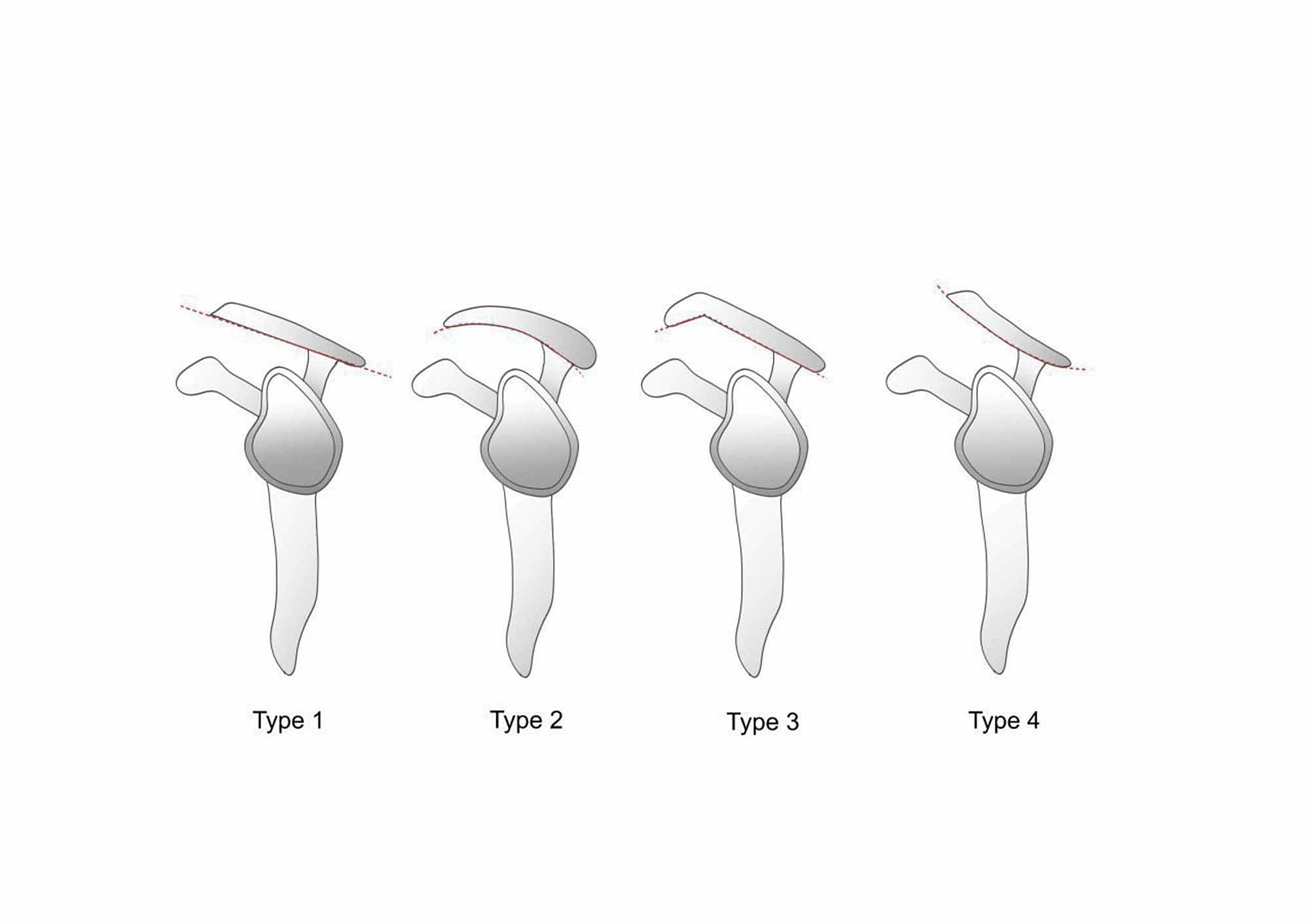

Ligamentous connection of the coracoacromial ligament and the rotator interval capsule is thought to prevent inferior migration of the glenohumeral joint. This structure provides an osseo-ligamentous static restraint to superior humeral head displacement. The coracoacromial ligament is a thick triangular ligamentous structure extending inferomedially from the anterolateral undersurface of the acromion to the lateral border of the coracoid process. Known as the “Surgeon’s Lighthouse,” the coracoid process serves as a landmark to avoid neurovascular injury, as major neurovascular structures traverse medially to the coracoid process. 10 The coracoid process can be palpated below and at the lateral edge of the clavicle. Also, the coracoid is an attachment site for the coracohumeral, coracoacromial, coracoclavicular, and superior transverse ligaments. The coracoid is an anchor point for several ligaments and tendons, including the pectoralis minor, coracobrachialis, and short head of the biceps brachii tendons. The coracoid process is a hook-like osseous structure arising from the superolateral edge of the scapula and extends in an anterolateral orientation.

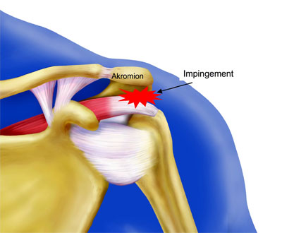
Processes that decrease the space within the coracoacromial arch presumably may lead to impingement-like symptoms. The humeral head provides the posterior/inferior border of the arch (Figure 1). Inferior to these structures, and coursing through the arch, are the subacromial/subdeltoid bursa, supraspinatus tendon, and biceps tendon. The coracoacromial arch is composed of (from anterior to posterior) the coracoid process, coracoacromial ligament, and the acromion process. The coracoacromial arch provides a safeguard for the shoulder, limiting superior migration of the humeral head. This knowledge will allow radiologists to effectively communicate with clinicians and help guide appropriate treatment. The purpose of this article is to help understand the relevant anatomy with regard to the subacromial region, review the imaging findings, and discuss the differing etiologies of extrinsic impingement. 3,9 Numerous operative and nonoperative treatment options have been described for extrinsic impingement, ranging from physical therapy and injections to acromioplasty and Mumford procedures.

8 Extrinsic shoulder impingement is most commonly related to mechanical compression from the acromion, acromioclavicular joint, and the coracoacromial ligament. 6,7Įxtrinsic, or external, impingement is one of the more common causes of shoulder pain and a frequent source for an orthopedic evaluation. The infraspinatus tendon is commonly injured, especially in patients under age 30, with MRI findings ranging from undersurface tears to complete tears. 6 MRI appearance of intrinsic impingement is varied and includes labral and rotator cuff pathology.

5 It is a result of extreme abduction and external rotation, which can lead to entrapment of the supraspinatus and/or the infraspinatus tendons between the glenoid. Intrinsic shoulder impingement is relatively uncommon and seen almost exclusively with overhead throwing athletes. 3,4 Two predominant types of shoulder impingement have been described: intrinsic and extrinsic. Rotator cuff impingement was first described by Neer et al when he stated that 95% of rotator cuff tears were attributed to impingement and generally occur in patients over age 40. 1,2 Rotator cuff pathology is a common etiology for shoulder pain, with impingement of the rotator cuff often playing an important role. Shoulder pain is a common musculoskeletal medical condition affecting 7% to 26% of individuals and is the third most common musculoskeletal-related complaint in the primary care setting.


 0 kommentar(er)
